3D imaging to decipher movement of malaria parasite
(Harvard Gazette, November 21, 2013)
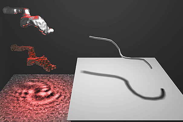 3D imaging has entered the laboratory as an important research tool. Researchers from Harvard have now employed this technique to produce detailed 3-D images of malaria sperm — the cells that reproduce inside infected mosquitoes — that shed new light on how the cells move. “The working assumption was that this structure moved through a consistent clockwise beating,” Laurence Wilson, a fellow at Harvard's Rowland Institute, “but what we found was, if you look at the malaria swimming, it doesn’t just move in a ‘right-handed’ way — it actually turns out to be a very general motor." Understanding how malaria parasites move may one day help scientists develop new strategies for combating the disease by halting its ability to reproduce.